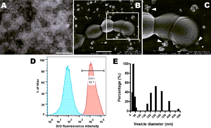
Fig 1. GBS produces extracellular membrane vesicles.
A. Representative transmission electron micrograph of membrane vesicles (MVs) isolated from GBS strain A909. Scale bar; 200 nm. B. Representative scanning electron microscopic image of GBS strain A909 exhibiting aggregated MVs dispersed around the cells and budding of MVs from GBS cells. Scale bar; 1 μm. C. Zoomed in view of the boxed area in (B). Arrowheads indicate vesicles budding sites. Scale bar; 100 nm. D. Flow cytometry of isolated MVs stained with lipid specific dye, DiO. Blue shaded curve denotes unlabeled MVs and pink shaded curve depicts DiO labeled MVs. E. Size distribution of isolated MVs by Dynamic Light Scattering. Experiments were performed thrice and data from one representative experiment is shown.

