Fig 7.
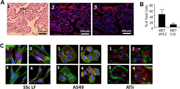
A. Expression of MET in lung tissues of patients with SSc-ILD. Panel 1 represents lung sections stained with hematoxylin and eosin (H&E). Panels 2 and 3 represent immunofluorescent images stained with anti-MET 4F8.2 antibody detecting total (cleaved and uncleaved MET) and anti-MET antibody C12 that does not recognize MET after C-terminal cleavage. Nuclei are stained with 4’,6-diamidino-2-phenylindole (DAPI). Representative images from three patients with SSc-ILD are presented. B. Quantitative results of image analysis for MET 4F8.2- and MET C12-positive cells. Cells (total, 4F8.2 positive and MET C12 positive) were counted on six randomly selected, no overlapping, high-power fields per sample at x400 magnification, and presented as mean and SD. C. Expression of endogenous MET in scleroderma lung fibroblasts (SSc LF), A549, and ATII cells. Cells were cultured in 4-chamber slides, challenged with (images 3 and 4) and without (images 1 and 2) cisplatin, fixed by 4% formaldehyde and stained with anti-MET 4F8.2 antibody (images 2 and 4) and anti-MET C12 antibody (images 1 and 3). Nuclei were stained with 4’,6-diamidino-2-phenylindole (DAPI). Phalloidin was used to show cytoskeleton.

