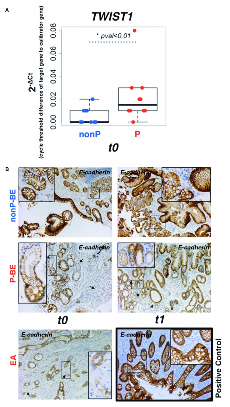
Fig 3. Changes in epithelial-to-mensenchymal biomarkers are visible in early and late BE backgrounds.

A. qRT-PCR validation of TWIST1 transcription factor in the patient-matched BE index biopsies, free of dysplasia/EA (t0). B. E-cadherin protein levels were evaluated by immunohistochemistry staining in P-BE associated with EA (t1) and in the patient-matched BE index biopsies, free of dysplasia/EA (t0). Arrows denote foci of lower E-cadherin expression. Normal appendix was used as E-cadherin immunostaining positive control. (Magnification: picture ×100, detail ×200).
