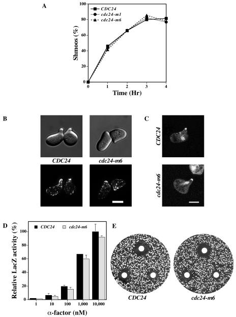
FIG. 3.
cdc24-m6 cells respond to mating pheromone. (A) cdc24-m6 cells form shmoos at the same rate and to the same extent as wild-type cells. Indicated strains were incubated with α-factor (12 μM) and shmoos were counted at the times shown (n = 125). The timing of the appearance of additional mating projections was the same in both strains. (B) The actin cytoskeleton in cdc24-m6 shmoos is polarized. DIC and fluorescence images of cells treated with 12 μM α-factor for 2 h as previously described in the legend to Fig. 2C are shown. Bar, 5 μm. (C) Cdc24-m6p localizes to mating projection tips and nuclei in shmoos. Confocal microscopy images of cdc24Δ cells expressing Cdc24-GFP or Cdc24-m6-GFP were treated with α-factor as described above. Bar, 5μm. (D) cdc24-m6 cells induce the mating-specific FUS1 gene in a pheromone-dependent fashion. Cells containing the FUS1-lacZ plasmid pSG231 were incubated with the indicated α-factor concentration for 1 h and LacZ activity was determined. The means of two independent experiments are shown with error bars indicating values. LacZ activity for CDC24 cells treated with 10,000 nM α-factor (40.8 Miller units) was set at 100%. (E) cdc24-m6 cells arrest growth in the presence of mating pheromone similar to wild-type cells. α-factor (1, 0.5, and 0.2 μg) was spotted on filters placed on a lawn of the indicated strain. Plates were incubated for 2 days. Measurements of the halo diameter indicated ≤5% difference between CDC24 and cdc24-m6 halos.

