
Figure 3.
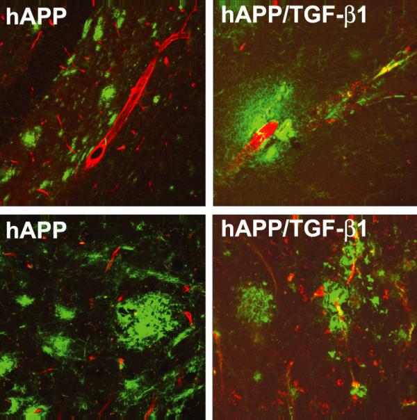
TGF-β1 overexpression in hAPP mice leads to CAA and reduces total brain amyloid. hAPP mice demonstrate amyloid plaques that are predominantly parenchymal (left panels), while bigenic hAPP/TGF-β1 mice (right panels) display fewer parenchymal amyloid plaques and have Aβ deposits localized to blood vessel walls (Aβ, green; Glut-1, red).