
Fig. 1.
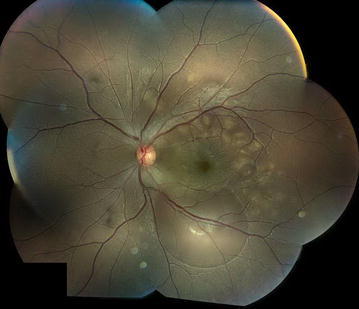
Left eye fundus photograph at the time of disease onset. Multiple, creamy yellow, flat, placoid lesions at the level of retinal pigment epithelium and multiple areas of retinal elevation consistent with sub-retinal fluid were visualized in the posterior pole