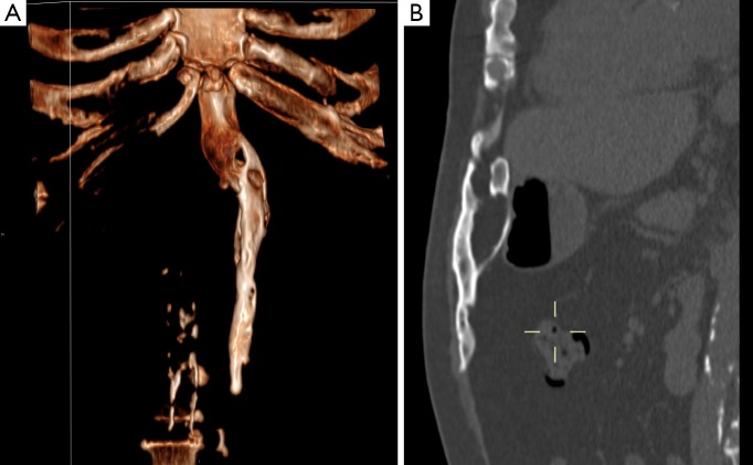
Figure 2.
A 53-year-old man with a symptomatic heterotopic ossification (HO) in an upper midline laparotomy scar. Protocol: CT-scan 75 mAs, 120 kV, 2 mm slice thickness. No contrast medium. (A) The sagittal plane reconstructions demonstrate the HO with in close relation to the xyphoid process extending caudally in the anterior abdominal wall. Periperal mineralization with central lucency is typical for mature HO; (B) an anterior oriented 3D volume rendered CT-image demonstrates the smooth surface of the HO, the relation with the xyphoid process and a slight bend to the left. In addition, the ribcage and spine are partial visible. The craniocaudal length is 18 cm.

