
Figure 2.
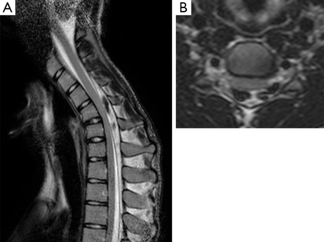
T2-weighted sequence on sagittal (A) and axial (B) planes. Posterior detachment and forward shifting of the dural sac with presence of epidural flow-voids from posterior venous plexus congestion, during neck-flexion (A). Antero-posterior cord flattening is evident on the axial plane (B).