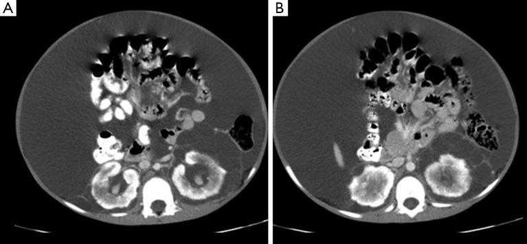
Figure 3.
Contrast enhanced CT (CECT) abdomen axial sections at the level of kidneys showing multiseptated collections seen involving bilateral perinephric and peripelvic space (left bigger than right) (A,B) with scalloping of right renal cortex (B) and gross ascites with central displacement of bowel loops.

