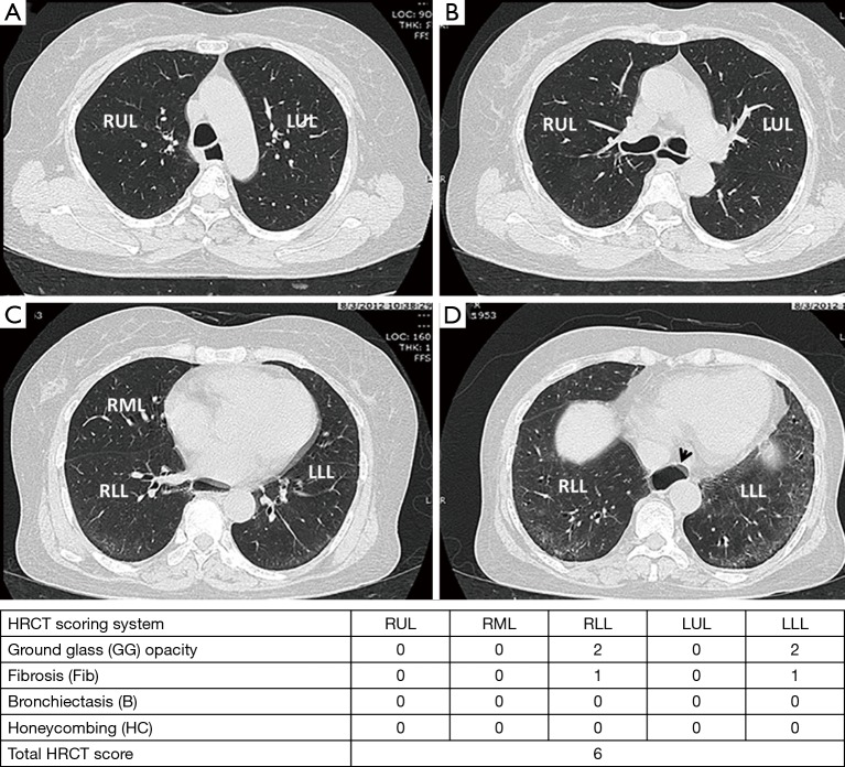
Figure 3.
Four high-resolution computed tomography (HRCT) images in a 58-year-old man with dcSSc at level of aortic arch (A); carina (B); inferior pulmonary vein (C) and lung base (D) reveals bilateral peripheral ground glass (GG) opacities with fine reticulation (Fib) and relative subpleural sparing, which compatible with early fibrotic nonspecific interstitial pneumonitis (NSIP) pattern. There was esophageal dilatation (arrow head).

