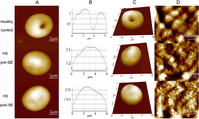
Fig. 2.

Representative AFM surface topographic images of erythrocytes from one healthy individual and one HS patient before and after splenectomy. a Single erythrocytes. b Height profile of the corresponding line in a. c 3D mode of single erythrocytes from a. d Surface ultrastructure on corresponding cells in images a. Scanning area: 10 μm × 10 μm in a, b, and c; 1 μm × 1 μm in d
