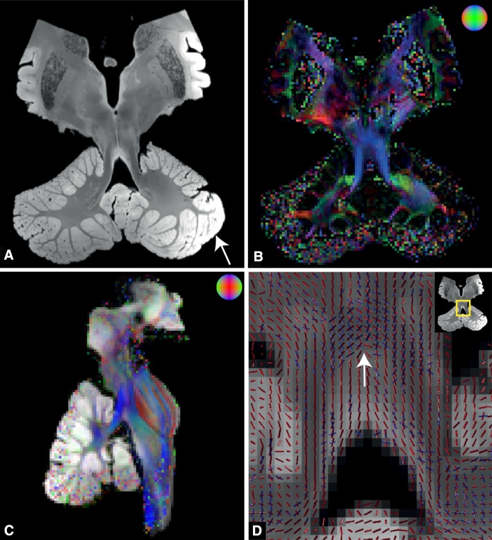
Fig. 1.
Postmortem MRI acquisitions. a Coronal view of the structural image acquired with the TRUFI sequence. White arrow inhomogeneous signal intensity in the cerebellar cortex. b Direction encoded colour (DEC) map with fractional anisotropy modulated intensity. Colour coding: green anterior–posterior, red left–right, blue inferior–superior. c The registration accuracy between structural and diffusion space is illustrated by overlaying DEC map with the structural MRI (sagittal view). The structural MRI was transformed to diffusion space with a 3D affine transformation. d ROI of diffusion directions in the decussation of the DRTTs (white arrow). Red and blue lines the first and second diffusion direction within a voxel, respectively

