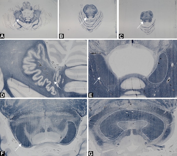
Fig. 3.
Histological axial sections stained for myelin with the modified Heidenhain–Woelke stain at different levels of the cerebellum and the pons. a The unstained dentate nucleus (white arrow) is clearly visible and characterized by its dented pattern. b The wedge-shaped structures represent the DRTTs (white arrow for left DRTT) in the superior cerebellar peduncles. c The decussation (white arrow) of left and right DRTTs at the level of the mesencephalon. d Close-up from a of the left dentate nucleus. The white matter enclosed by the dentate nucleus forms the origin of the DRTT. e Close-up of the DRTT within the superior cerebellar peduncles. Densely packed white matter is surrounding the DRTT at this level. Difference in texture allowed for differentiation of the DRTT with adjacent white matter indicated by the white arrows. The dashed outline marks the right DRTT in this section. f Close-up from b of the DRTT (white arrow and outline, for left and right DRTT, respectively) within the superior cerebellar peduncle, but more superior located as in (e). g Close-up from c of the decussation (white outline) of the superior cerebellar peduncles in the mesencephalon

