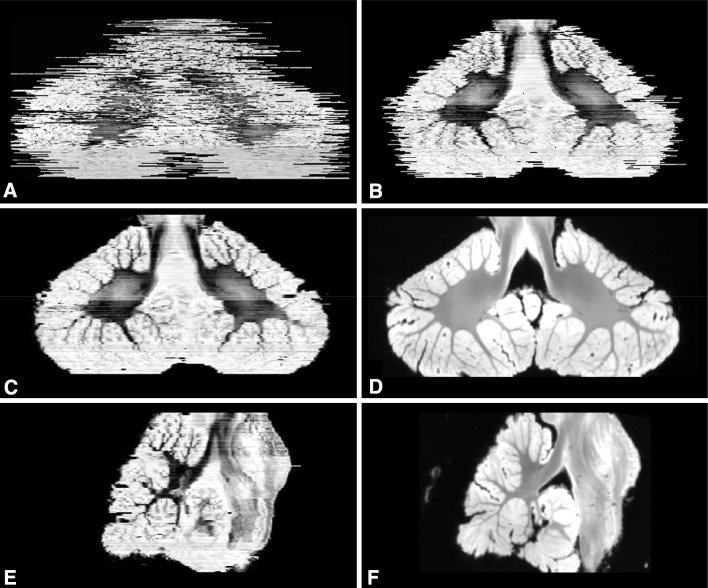
Fig. 4.
3D reconstruction of the histological sections from the cerebellum and brainstem. a, b Coronal views from before and after affine registration of the stacked slices, respectively. Following affine registration, a non-linear registration approach was applied and represented in a coronal (c) and sagittal view (e). Alignment of internal structures significantly improved after this step and was most pronounced in white matter. Corresponding MRI slices for c and e are depicted in d and f, respectively

