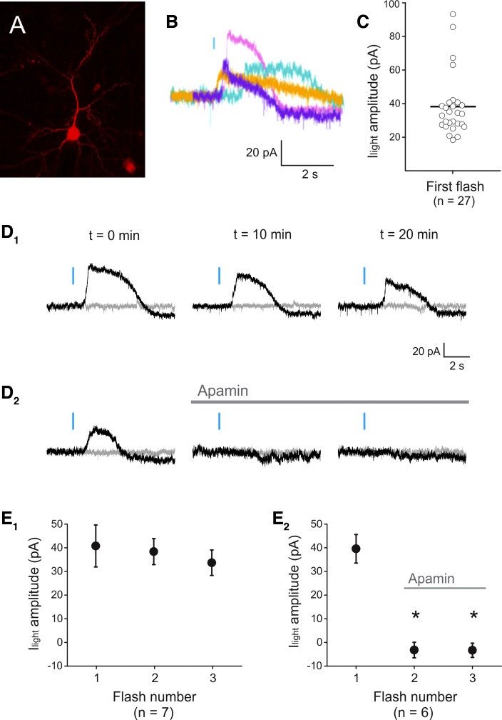
Fig. 2.
Light-induced activation of a phasic outward current in melanopsin-expressing pyramidal neurons. A: image derived from a flattened confocal z stack depicting a melanopsin-expressing pyramidal neuron identified by coexpression of a red fluorescent protein. B: light-evoked transient outward currents recorded in 4 different pyramidal cells. Blue bar denotes the light flash. C: graph plotting the amplitude of the light-evoked transient outward current (Ilight) amplitude observed in the present experiments. D1: light flashes delivered every 10 min could repeatedly evoke a transient outward current in a single pyramidal cell. D2: these light-evoked transient outward currents were blocked by administration of apamin (100 nM). Gray traces illustrate a sweep without light, collected immediately before the light sweep. E1: graph illustrating the relative stability of the light-evoked transient outward currents upon repeated light stimulation in 7 cells (P = 0.756, ANOVA). E2: graph summarizing the effect of apamin (100 nM) on the light-evoked transient outward currents (P = 3.48E-6, ANOVA, n = 6 cells). *Statistically significant inhibition.

