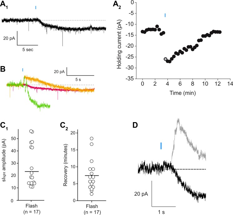
Fig. 4.
Light-evoked sustained inward current in melanopsin-expressing pyramidal cells. A1: under voltage clamp a 20-ms flash of blue light causes the appearance of a slowly developing inward current. A2: graph plotting the time course for the light evoked inward current. Gray circle corresponds to the trace illustrated in A1. B: overlapped traces illustrating the variability in the early time course of the light-evoked resulting from the summation of variable light-induced transient outward and sustained inward currents in 3 cells. C1: graph plotting the amplitude of the light-induced inward current (sIlight) recorded in the present experiments. C2: graph plotting the recovery time for the light-induced inward current in the same 14 cells. D: overlapped traces depicting the light-evoked current observed under control conditions (gray trace) and after bath application of apamin (100 nM, black trace). Blockade of the transient outward current by apamin revealed the time course of the sustained inward current.

