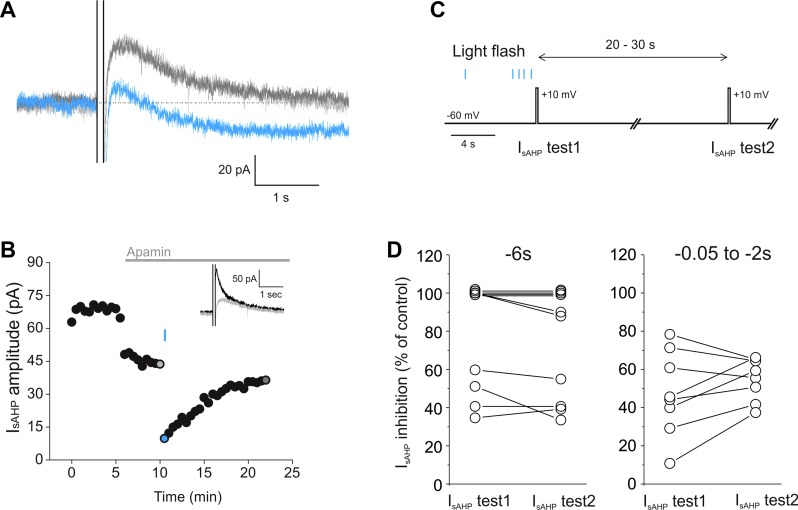
Fig. 5.
Light-induced inhibition of the IsAHP in melanopsin-expressing cortical pyramidal cells. A: in voltage clamp, a depolarizing pulse (+30 mV, 100 ms) induced the appearance of a IsAHP (light gray trace) in a melanopsin-expressing pyramidal neuron. A brief (20 ms) flash of light caused prolonged inhibition of the IsAHP (blue trace), which recovered over time (dark gray trace). B: graph plotting the light-induced inhibition of the IsAHP in the cell pictured in A with colored circles corresponding to the individual traces. As shown in the inset, the IsAHP was initially isolated by bath administration of apamin (300 nM). (black trace is prior to apamin; light gray trace is after apamin and corresponds to the same color trace shown in A). C: 2-pulse protocol used to obtain information regarding the time course of the onset of the light-evoked IsAHP inhibition. D: when a brief flash of light was administered 6 s prior to the depolarizing pulse used to evoke IsAHP (−6 s), the extent of IsAHP inhibition (IsAHP test 1) (69.5 ± 11.2%) was not significantly different from that observed 20–30 s later (IsAHP test 2) (65.2 ± 11.3%) (n = 7, P = 0.79, Student's t-test). The extent of IsAHP inhibition was also not significantly different between IsAHP test 1 and IsAHP test 2 when the light stimulation for IsAHP test 1 occurred between −2 and −0.05 s prior to the pulse (47.5 ± 4% compared with 55.1 ± 8%, n = 8, P = 0.4, Student's t-test). For this analysis the amplitude of the IsAHP was taken as the mean of a 100-ms segment beginning ∼350 ms after the end of the depolarizing step and was baseline-subtracted.

