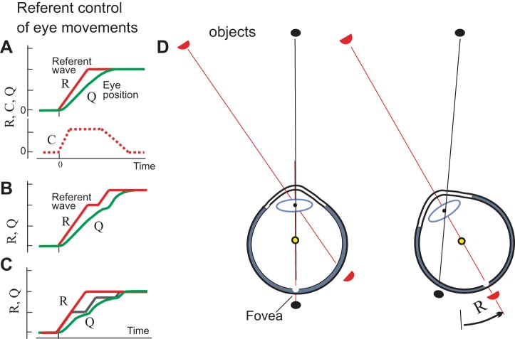
Fig. 7.
Referent control of eye motion and visual space constancy. A: saccadic motion results from appropriate shifts in the tension-extension characteristic of EOMs shown in Fig. 6. As in arm muscles, these shifts result from a combination of referent reciprocal (R) and referent coactivation (C) commands, both producing changes in the eye threshold position at which EOMs begin to generate active tension. When an object of interest is defined, saccadic referent eye rotation is initiated that virtually brings the object image to the fovea. Actual eye rotation (Q) follows. Q lags behind the changes in R because of eye inertia, gradual muscle activation and force generation as well as of passive resistance of connective tissue in the orbit. Because of transient discrepancy in R and Q, to avoid misperception, vision is blocked until the eyes come to the final referent position. The C command contributes to saccade speed and can be minimized in pursuit. Shifts in R can be associated with wave propagation along a neural ensemble (referent wave). EOMs generate activity depending on the difference between the actual and the referent position of the eyes. B: if necessary, a second saccade can be initiated even before the end of the first saccade. C: the R can predetermine a total shift in gaze, but at a lower, executive level this shift can be produced in several saccadic steps. D: referent eye rotation (R) symbolizes transition to a new spatial frame of reference in which retinal images are considered such that the external world is perceived as stationary despite retinal shifts in its images. [D reproduced from Feldman 2015 with permission from Springer. Copyrights Springer.]

