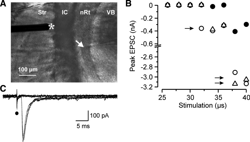
Fig. 1.
Minimum stimulation protocol. A: photomicrograph of an acute horizontal thalamic slice with the recording pipette (arrow) located in the thalamic reticular nucleus (nRt) and the stimulating electrode (*) positioned in the striatum near the internal capsule (IC) where it activates thalamocortical (TC) and corticothalamic (CT) axonal projections. Str, striatum; VB, ventrobasal complex. B: peak amplitude of inward excitatory postsynaptic currents (EPSCs) recorded in a nRt cell at a holding potential of −70 mV during a minimum stimulation paradigm. The intensity of the stimulus was initially fixed slightly above threshold for a 50-μs stimulus and the duration was then progressively stepped from in 2-μs steps from a subthreshold stimulus (25 μs, all failures) until all-or-none responses were triggered. Three consecutive duration series, each represented by a different symbol, of the stimulus were repeated. The minimal stimulation duration was selected as that which was in the middle of the range of durations that evoked threshold responses (as defined as intermixed failures and successes, with the latter being of relatively fixed amplitude, marked with a single arrow). Higher stimulus intensities (longer duration) led to a recruitment of additional fibers and larger responses (double arrows). C: current responses from one trial of the minimum stimulation paradigm (26–36-μs durations shown, corresponding only to either failures or minimal responses shown as solid circles in B). Note that the onset of the unitary EPSCs occurs at a fixed latency after the stimulation artifact (solid circle) and their time to peak and amplitude remain stable.

