
FIGURE 9.
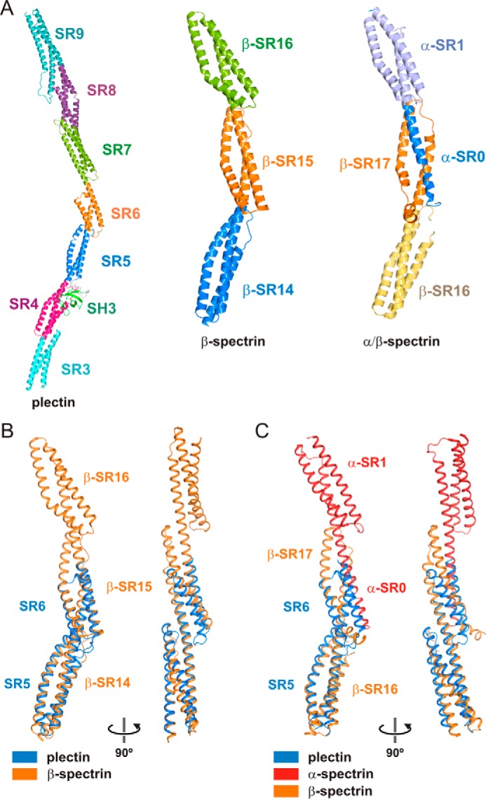
Comparison of the plakin domain of plectin with curved multi-repeat segments of spectrins. A, ribbon representation of the composite structure of the plakin domain of plectin, the crystal structure SR14–SR16 of human β2-spectrin (PDB code 3EDV), and the tetramerization domain complex of human erythroid spectrins formed by the partial SR0 and SR1 of α-spectrin and the SR6 and the partial SR17 of β-spectrin (PDB code 3LBX). B and C, superimposition of the structure of the SR5-SR6-ΔSH3-A of plectin onto repeats SR14-SR15 of β2-spectrin (B) or repeats β-SR16-β-SR17/α-SR0 of the tetramerization complex of α/β spectrins (C).