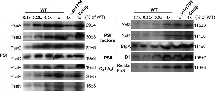
FIGURE 6.
Representative membrane protein profiles in WT, the Δslr1796 mutant, and the complemented strain. Immunoblotting analyses of photosynthetic membrane proteins are shown. Equal amounts of membrane proteins were loaded for the detection of each protein. A dilution series of WT protein (10, 25, 50, and 100%) were loaded. Antibodies were directed against PSI subunits (PsaA, PsaB, PsaC, PsaD, PsaE, PsaF, and PsaK), PSI assembly factors (BtpA, Ycf3, and Ycf4), PSII core subunit D1, and a subunit of the cytochrome (Cyt) b6f complex (Rieske FeS). The number at the right of each band is the percentage of protein in the Δslr1796 mutant relative to that of WT (set as 100%). Values represent the standard deviation of at least 10 measurements for each protein.

