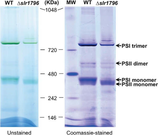
FIGURE 7.

Blue native PAGE of membrane protein complexes of WT and the Δslr1796 mutant. An equal protein amount of about 35 μg was loaded to each lane. The gel is shown as unstained (left) and stained with Coomassie Brilliant Blue (right). Molecular mass markers are indicated, and the assignments of PSI and PSII complexes are given.
