FIGURE 1.
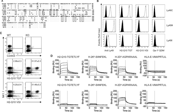
H2-Q10 is a bone-fide ligand for C57BL/6 Ly49C. A, sequence alignment of H2-Q10, H-2Kb, H-2Dd, and Qa-1b. Residues conserved in at least three of the sequences are shown as dots. Binding sites for Ly49C on H-2Kb are shown within boxes. B, tetramer staining of CHO cells expressing Ly49C, Ly49H, and Ly49I. Open curves show staining with Ly49-specific antibody (Anti Ly49), H2-Q10 loaded with TGTETLYF (H2-Q10 TGT), H2-Q10 loaded with VGITNVDL (H2-Q10 VGI), or Qa-1b loaded with AMAPRTLLL (Qa-1b QDM); black filled curves show isotype control antibody (for anti-Ly49) or irrelevant human HLA-E tetramer (for H2-Q10). Data are representative of at least 5 independent experiments. C, H2-Q10 binding to Ly49C ex vivo is impaired by cis interactions with MHC-I. Splenocytes were prepared from WT and H-2Kb/Db double-deficient mice. Cells were stained with NK1.1, CD3, an irrelevant human HLA-E tetramer (control), H2-Q10 tetramer loaded with TGTETLYF (H2-Q10 TGT), or H2-Q10 loaded with VGITNVDL (H2-Q10 VGI). The dot plots show events that have been electronically gated as singlets, live and CD3− (not shown), and presented as NK1.1 (vertical) versus tetramer (horizontal). Numbers in the top right quadrant of the H2-Q10-stained panels represent the percentage of H2-Q10+ NK cells and are presented as mean ± S.E. of four mice. Data are representative of 2 independent experiments using four mice per experiment (n = 8). D, Ly49C binds with similar affinity to H2-Q10, H-2Kb, and H-2Dd. Binding decreasing concentrations (30, 12, 4.8, 1.92, 0.77 μm; solid lines top to bottom) of Ly49C (upper) or Ly49A (lower) to streptavidin-immobilized H2-Q10-TGTETLYF, H-2Kb-SIINFEKL, H-2Dd-AGPARAAAL, or HLA-E-VMAPRTLIL is shown. Results are presented in response units (RU) after subtraction of the baseline values. Solid vertical lines at 0 and 60 s indicate injection start and stop, respectively. Binding affinity (determined by equilibrium analysis): H2-Q10-TGTETLYF-Ly49C KD = 5.0 ± 0.5 μm; H-2Kb-SIINFEKL-Ly49C, KD = 1.0 ± 0.2 μm; H2-Dd-AGAPARAAAL-Ly49C, KD = 1.3 ± 0.5 μm; H2-Dd-AGAPARAAAL-Ly49A KD = 0.5 ± 0.1 μm. Responses for HLA-E with Ly49A and Ly49C, H2-Q10 with Ly49A, and H-2Kb with Ly49A are below the limit of detection.

