FIGURE 2.
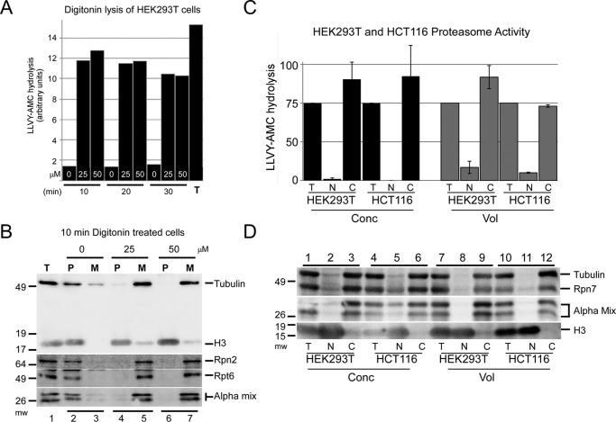
Proteasome peptidase activity can be released into the medium. A, HEK293T cells were treated with varying concentrations of digitonin and pelleted. Following digitonin treatment, the peptidase activity that was released into the medium (M) was compared with that detected in the cell pellet (P). Prolonged exposure to digitonin (30 min) did not further increase peptidase activity in the medium. Total peptidase activity (T) was determined by lysing untreated cells and measuring LLVY-AMC hydrolysis. B, HEK293T cells were treated for 10 min with varying concentrations of digitonin (0, 25, and 50 μm), and immunoblotting was used to determine the proteasome subunit levels in the medium (M) and cell pellet (P). The detection of tubulin and histone H3 verified the location of a cytosolic and nuclear protein, respectively. Basal levels of Rpn2, Rpt6, and α-proteasome subunits were established using lysates prepared from untreated cells (lane 1). Histone H3 was recovered in the pellet in both treated and untreated cells (lanes 2, 4, and 6). Treatment with digitonin resulted in significant depletion of proteasome subunits from the pellet fraction (lanes 4 and 6) and their recovery in the medium (M, lanes 5 and 7). C, nuclear and cytosolic fractions were prepared from HEK293T and HCT116 cells. LLVY-AMC hydrolysis was measured in the fractionated lysates using either an equal amount of protein (Conc) or proportional volumes (Vol). Peptidase activity in both cell lines was predominantly cytosolic. The sum of cytosolic (C) activity was similar to total (T) activity, indicating that the proteasome activity is predominantly in the cytosol. D, protein extracts described in C were examined by immunoblotting. Rpn7 and multiple α-subunits (Alpha mix), as well as tubulin, were detected primarily in the cytosol (C) in both cell lines, whereas histone H3 was found in the nucleus (N). Analysis of equal protein (Conc) significantly over-represents the nuclear fraction, as evidenced by the high levels of histone H3 in the nuclear (N) fraction when an equal amount of protein (Conc) was examined. In contrast, proteasome subunits were not detected in the nuclear fraction when we examined proportional volumes of the fractionated lysates. Similar findings were observed in two independent cell lines (HEK293T and HCT116).

