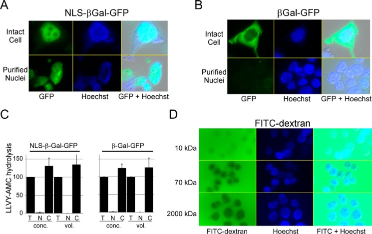
FIGURE 4.
Purified nuclei are structurally intact. A, HEK293T cells were transfected with a vector expressing NLS-βGal-GFP, and fluorescence was detected predominantly in the nucleus (upper panels). Following fractionation, GFP fluorescence was retained in the purified nuclei (lower panels). B, βGal-GFP (lacking a nuclear localization signal) was similarly expressed in HEK293T cells, and cytosolic expression was observed (upper panels). Purified nuclei showed no evidence for GFP fluorescence (lower panels). C, we measured proteasome peptidase activity in cells described above. LLVY-AMC hydrolysis in the cytosol (C) and nucleus (N) was compared with activity present in total lysate (T). Error bars indicated S.E. D, nuclei were isolated from HEK293T cells and incubated with FITC-dextran in the sizes indicated. FITC 10-kDa dextran readily entered the nucleus, whereas FITC 2,000-kDa dextran was entirely excluded. Hoechst staining of nuclei and a merged differential interference contrast image are also shown.

