
FIGURE 1.
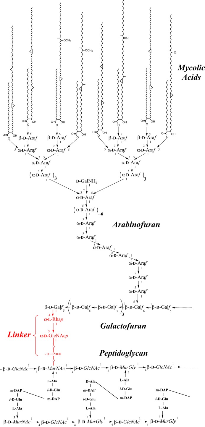
The M. tuberculosis cell wall core. PG from M. tuberculosis is composed of linear chains of N-acetyl-α-d-glucosamine and modified muramic acid substituted with peptide side chains that are heavily cross-linked (70–80%), providing added structural integrity to the bacterium. AG is attached to PG through a phosphodiester link to position 6 of some of the Mur residues. The specific linker unit ensuring its covalent attachment to PG is shown in red. One arabinan chain is shown here attached to the galactan domain. The characteristic Ara6 non-reducing termini of the arabinan domain of AG serve as the anchoring points for the mycolates. Only one arabinan chain is shown for clarity.