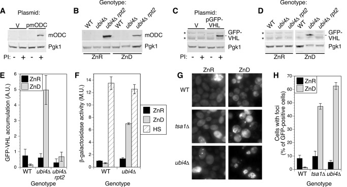
FIGURE 5.
Reduced proteasome activity restores ubiquitin-dependent degradation. A, mODC accumulates following proteasome inhibition (PI). BY4743 pdr5Δ cells transformed with empty vector (pRS315; V) or p415-GFP-FLAG-mODC (pmODC; expressing FLAG-tagged mouse ornithine decarboxylase) were grown to log phase in LZM + 100 μm ZnCl2 and treated for 1 h with DMSO carrier (−) or proteasome inhibitors (50 μm MG132 and 250 μm bortezomib; +) before harvesting. Protein extracts were analyzed by immunoblotting with anti-FLAG antibodies. B, mODC accumulates in rpt2E301K mutants independently of zinc supply. Wild-type (BY4741), ubi4::KanMX4, and ubi4Δ rpt2E301K (CWM280) strains transformed with p415-GFP-FLAG-mODC were grown to log phase in LZM + 100 μm ZnCl2 (ZnR) or 1 μm ZnCl2 (ZnD) prior to immunoblotting. C, GFP-VHL accumulates following proteasome inhibition. BY4743 pdr5Δ was transformed with empty vector (V) or pESC-LEU-GAL1p-GFP-VHL (pGFP-VHL; expressing GFP-tagged von Hippel-Lindau protein), and cells were assayed for mODC as described in A. The position of the GFP-VHL band is shown, and two nonspecific bands are indicated by asterisks. D, GFP-VHL is stabilized in zinc-deficient ubi4Δ cells and degraded in ubi4Δ rpt2E301K. GFP-VHL accumulation was assayed as described for mODC in B. For all immunoblots in A–D, one blot representative of three replicates is shown, and Pgk1 was detected as a loading control. E, quantitation of GFP-VHL bands for three replicates, including the blot shown in D. Error bars denote ±1 S.D. F, expression of an Hsf1-regulated reporter gene in wild-type and ubi4Δ cells. Strains transformed with the pHSE-lacZ plasmid were grown to log phase in zinc-replete (ZnR; LZM + 100 μm ZnCl2) or zinc-deficient (ZnD; LZM + 1 μm ZnCl2) medium and assayed for β-galactosidase activity. As a positive control for reporter activity, aliquots of zinc-replete cells were also subjected to heat shock at 37 °C for 1 h before assay (HS). G and H, wild-type (BY4743), tsa1Δ, and ubi4Δ diploid strains transformed with pHSP104-GFP were grown in LZM + 1000 μm ZnCl2 (ZnR) or 1 μm ZnCl2 (ZnD) medium for at least four generations, maintaining cell density below an A595 of 0.4 by dilution with fresh media. Cells were examined by fluorescence microscopy to determine the proportion of cells with detectable GFP fluorescence that also displayed foci. Data points indicate the average of three replicates, and error bars show ±1 S.D. A.U., arbitrary units; M.U., Miller units.

