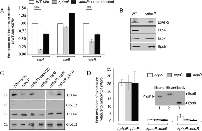
FIGURE 1.
PhoP-mediated espACD activation restores ESX-1-dependent ESAT-6 secretion of M. tuberculosis. A, quantitative RT-PCR to examine expression levels of espA, espB, and espR in ΔphoP and ΔphoP-complemented strains relative to WT M. tuberculosis H37Rv. The results represent average values with standard deviations derived from at least three independent RNA preparations (*, p < 0.05; **, p < 0.01; ***, p < 0.001). B, immunoblot analyses of 20 μg of cell extracts of WT and ΔphoP using appropriate antibodies. RpoB was used as a loading control. C, immunoblot analyses of 20 μg of culture filtrates (CF) or cell lysates (CL) of indicated M. tuberculosis strains. αGroEL2 was used as a control to verify cytolysis of cells. Note that complementation of ΔphoP M. tuberculosis with phoP, espB, espR, and espACD was carried out using p19Kpro as the expression vector and compared with ΔphoP M. tuberculosis carrying p19Kpro as the empty vector control (see “Experimental Procedures”). D, expression of espA, espC, and espD in indicated mutant M. tuberculosisH37Rv strains relative to ΔphoP-p19kpro (empty vector control) as measured by RT-PCR analysis. Inset shows ectopic expression of proteins as detected by immunoblotting with anti-His antibody. IB, immunoblot.

