FIGURE 3.
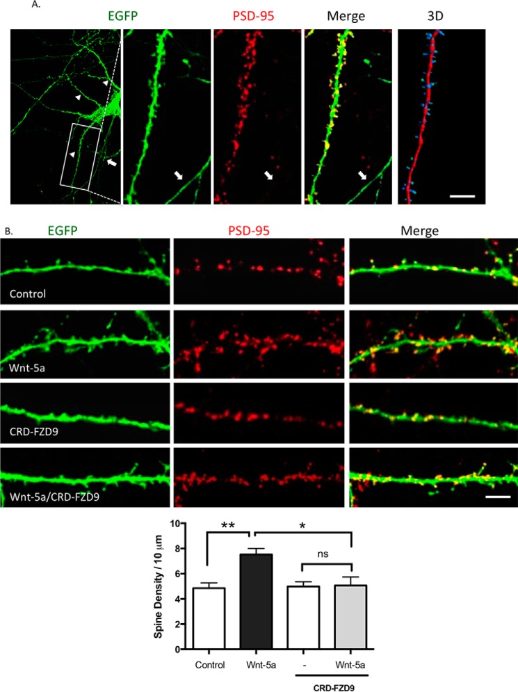
CRD-FZD9 inhibits the spinogenesis triggered by Wnt-5a treatment. A, left, immunodetection of PSD-95 (red) in EFGP-transfected neurons (green) maintained for 14 DIV. The Merge image shows the EGFP protrusion with the PSD-95 puncta stain in the head, indicating that the protrusions are spines. Arrowheads indicate dendrites, and arrows indicate an axon. Right, three-dimensional reconstruction of the dendrite. Scale bar, 25 μm. B, upper panel, representative images of EGFP-transfected neurons at DIV 10 and then treated with Wnt-5a, CRD-FZD9, or Wnt-5a+CRD-FZD9 for 2 h at DIV 14. Lower panel, quantification of spine density. Scale bar, 5 μm. (n = 3; 10 neurons/condition, 2–3 neurites/neuron). **, p < 0.01; *, p < 0.05.

