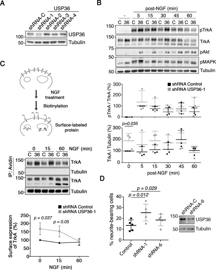
FIGURE 2.
USP36 as a regulator of TrkA levels, downstream signaling, and function. A, endogenous USP36 levels are reduced by shRNA-1. Western blotting showing USP36 levels in PC12-6/15 cells infected with lentivirus expressing control or USP36 shRNAs (shRNA-1–4) corresponding to the sequences of siRNAs present in the library used to perform the screening. B, USP36 protein regulates TrkA levels and the activation of the receptor and downstream signaling pathways in response to NGF. PC12-6/15 cells were infected with lentivirus expressing control shRNA or USP36 shRNA-1, and 5–7 days after infection, cells were treated as described in Fig. 1A. Active TrkA (pTrkA), Akt (pAkt), MAPK (pMAPK), and TrkA were detected using Western blotting. Tubulin was used as a loading control. A representative experiment is shown. Quantification of TrkA and pTrkA levels (bottom panels) is shown (means ± S.D.; n = 3; two-tailed unpaired Student's t test). C, a schematic diagram of the surface expression assay using the biotinylation procedure in response to NGF is shown (top panel). PC12 cells were infected with control and USP36 shRNA-1 lentivirus. The cells were treated with NGF as described in Fig. 1A and then biotinylated as described under “Experimental Procedures.” The cell lysates were prepared, surface proteins were subjected to precipitation with neutroavidin-agarose, and Western blotting analysis were performed with the corresponding antibodies (middle panel). Quantification of surface TrkA upon NGF treatment (means ± S.D.; n = 3, two-tailed unpaired Student's t test) is shown (bottom panel). D, PC12 cell differentiation in response to NGF is enhanced upon USP36 depletion. PC12 cells transfected with plasmids encoding for GFP and control shRNA, USP36 shRNA-1, or USP36 shRNA-6 were stimulated 2 days later with NGF (10 ng/ml) in serum-reduced medium to differentiate the cells. Western blotting analysis showing USP36 reduction in PC12 cells expressing USP36 shRNA-6 is shown (top panel). GFP-positive cells were scored for differentiation 72 h after the addition of NGF. The results are the means ± S.D. of five independent experiments counting at least 330 cells/condition (two-tailed unpaired Student's t test).

