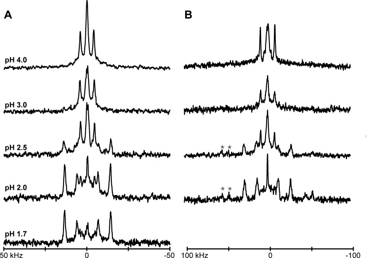
FIGURE 3.
Selected deuterium NMR spectra for two labeled alanines (7 and 9) in GWALP23-H12 in DOPC bilayers, hydrated with 10 mm buffer at the indicated pH, showing (A) β = 90° and (B) β = 0° sample orientations. The difference in spectra from sharp, well resolved signals above pH 4, to spectra with multiple signals below pH 2.5, indicates that the His12 residue is charged only under strongly acidic conditions. Red stars in B indicate signals with large |Δνq|, which likely correspond to backbone Cα-D nuclei of labeled alanine residues. The peptide/lipid ratio is 1/60 at a temperature of 50 °C.

