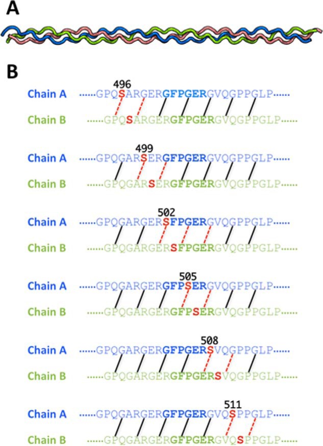
FIGURE 6.

A, the triple helix structure of the wild-type collagen peptide generated with the Triple-Helical Collagen Building Script (THeBuScr) (47). Chains A, B, and C are colored in blue, green, and red, respectively. B, interchain backbone NH···CO interactions are illustrated. Different backbone NH···CO interactions are disrupted in the mutants depending on the location of the substitution. Backbone hydrogen bonds whose average N···O distances were ≥3.5 Å during the course of simulation are labeled with red dashed lines to indicate their disruption. The hydrogen bonding is shown only for chains A and B of the triple helix because these are the chains that interact with integrin (23).
