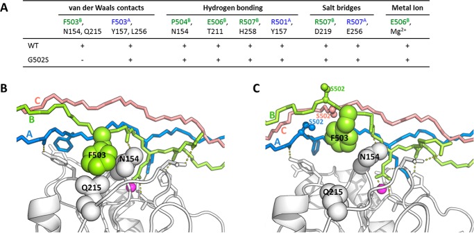
FIGURE 7.
A, the key interactions that participate in the integrin-collagen binding and the residues involved in each interaction. The residues from integrin and chains A and B of collagen are colored in black, green, and blue, respectively. All these interactions are observed to be stable in the 100-ns MD simulation of integrin with WT collagen (indicated by + symbols). The van der Waals interaction of collagen Phe-503B with integrin Gln-215 and Asn-154 is disrupted by the Gly to Ser mutations at position 502 (indicated by the − symbol). B and C, the simulation structures of integrin with WT collagen (B) and integrin with G502S mutant (C). Chains A, B, and C for collagens are colored in blue, green, and red, respectively. The Mg2+ ions are represented by magenta spheres. The substituted Ser residues are shown in ball-and-stick. The disrupted van der Waals interaction is illustrated.

