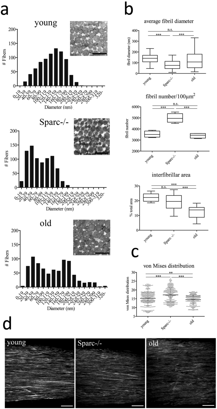
Figure 3. Structural analysis of healthy-aged and Sparc−/− C57BL/6 mouse Achilles tendons.
(a) Ultrastructural analysis of mouse Achilles tendons. Shown are frequencies of fibril diameters of young, Sparc−/− and healthy-aged C57BL/6 mouse determined by TEM (n = 800 fibrils of 3 animals each). Representative images are shown in the inserts. Mean ± SEM are displayed; Scale bar: 500 nm. (b) The average fibril diameters are significantly different between young and Sparc−/− tendons, young and healthy-aged tendons and Sparc−/− and healthy-aged tendons. Box plots represent measurements of a total of 800 fibrils; ***P < 0.001, **P < 0.01, non-parametric repeated Measures ANOVA (Friedman Test with Dunn’s post hoc test). Fibril number per area and interfibrillar area was determined for a total of 4 animals. (c,d) Second-harmonic generation microscopy and angle analysis of Collagen fibres in young, Sparc−/− and healthy-aged Achilles tendons. Dot blots represent von Mises distributions determined from z-stacks acquired from a total of 10 Achilles tendons per group. ***P < 0.001, **P < 0.01, One-way ANOVA (Dunn’s Multiple Comparison Test); Scale bars: 50 μm.

