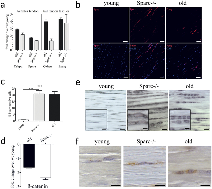
Figure 6. Increased lipid accretion and adipogenic marker expression in aged tendons.
(a) mRNA levels for the adipogenic marker genes Pparγ and Cebpα were determined by quantitative RT-PCR. Shown are fold changes over mRNA levels quantified for young Achilles tendons (left) and tail tendon fascicles (right). Data represent means ± SEM of 3 independent biological replicates. (b,c) Representative immunofluorescence microscopy images and quantification of Pparγ-positive cells within the dense connective tissue of young, healthy-aged, and Sparc−/− Achilles tendons. Graphs represent mean ± SEM of percent positive cells determined for each group (n ≥ 1000 cells counted from 3 Achilles tendon samples). Scale bars: 50 μm; ***P < 0.001, One-way ANOVA (Tukey’s Multiple Comparison Test). (d) β-catenin gene expression in Achilles tendon tissue. Shown is fold-change in expression compared to young tendon tissue. Bars represent mean ± SEM (n = 5). (e) Sudan Black B staining demonstrating accumulation of lipid droplets and change in cell morphology in Sparc−/− and healthy-aged Achilles tendons. Scale bars: overview: 20 μm; insert: 10 μm; (f) Immunohistochemical detection of the lipid droplet membrane protein Perilipin-1 in young, Sparc−/−, and healthy-aged tendons. Scale bars: 10 μm.

