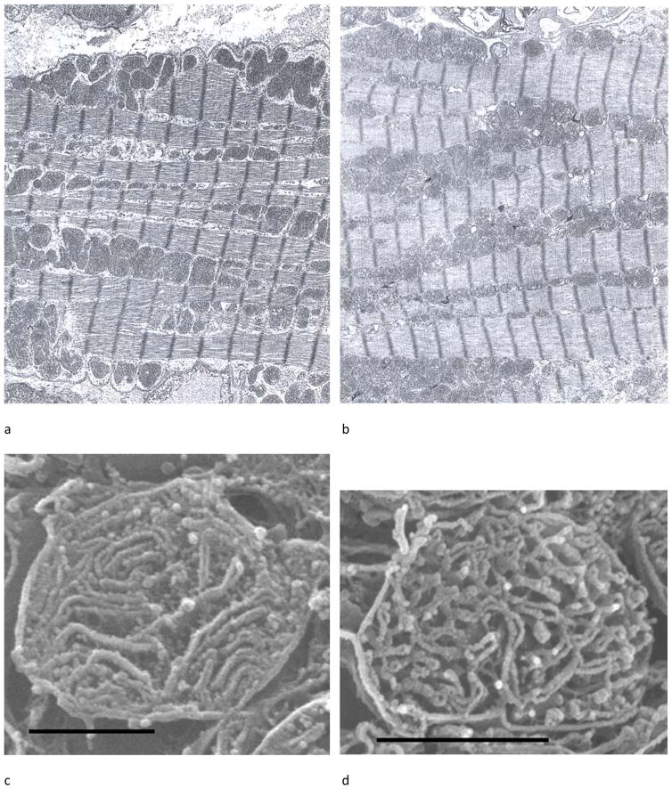
Figure 5.
(a) Osmium-extracted cardiomyocyte in situ from an adult rat heart observed in cross-section by HRSEM. The sarcolemma is pointed out by the series of white arrows. The SSM are under the sarcolemma; the black arrow to Fig. 1b where a typical SSM is shown at higher magnification. The box identifies the more central area where the myofibrils have been extracted by the osmium treatment exposing the IFM. The arrow to Fig. 1c shows an IFM in higher magnification. (b) A SSM containing lamelliform cristae exclusively. (c) An IFM with tubular cristae forming a lattice. Scale line for (a) = 4 μm and for (b) and (c) = 0.5 μm.
The images are taken from Figure 1a and Figure 2a and 2d from reference 46.

