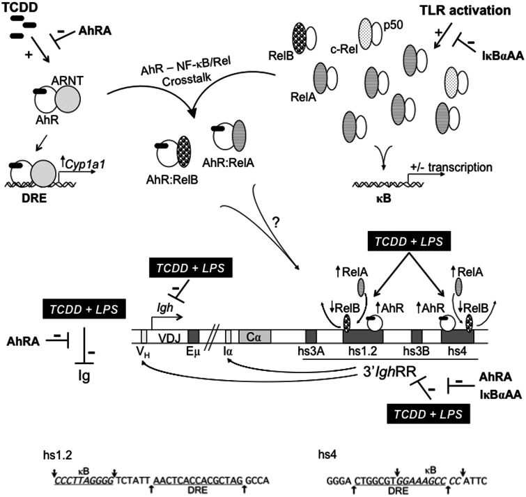
FIG. 10.
Schematic representation of mouse Igh regulation by the AhR and NF-κB/Rel proteins. A schematic depicting a portion of the mouse Igh gene locus including the VDJ antigen recognition region, the 3′ most Igh constant region Cα, which will encode for the heavy chain of IgA, and the regulatory elements: variable Igh promoter (VH), µ or intronic enhancer (Eµ), intronic promoter for Cα (Iα), and the 3′Igh regulatory region (3′IghRR) with its 4 enhancer regions (ie, hypersensitive sites [hs] hs3A, hs1.2, hs3B, and hs4). Long-range interactions between the 3′IghRR and the VH promoter and the intronic promoters just upstream of each constant region are noted with arrows from the 3′IghRR to these sites (Birshtein, 2014). The AhR and NF-κB/Rel nuclear pathways are depicted and show (1) TCDD-induced AhR activation and the prototypical response of Cyp1a1 induction; (2) toll-like receptor activation of NF-κB/Rel proteins, which can form various homo- or heterodimers and upregulate or downregulate the expression of various genes; and (3) crosstalk between the AhR and NF-κB/Rel proteins. The typical NF-κB/Rel heterodimers following IκBα degradation are depicted and will largely be RelA-p50 and less so of c-Rel-p50. Excess RelB not bound by inactivated, unprocessed p100 can be sequestered by IκBs, including IκBα (inhibitor kappa B-alpha protein) (Millet et al., 2013). The potential interactions between the AhR and the NF-κB/Rel proteins RelA and RelB are shown, which may account for the changes in RelA versus RelB binding within the hs1.2 and hs4 enhancers following TCDD and LPS cotreatment. The inhibitory effects of TCDD on LPS-induced 3′IghRR activation, Igh expression, and Ig secretion are depicted by( ). The inhibitory effects of the AhR antagonist (AhRA) and the IκBαAA superrepressor are also illustrated. Nucleotide sequences for the κB (NF-κB/Rel DNA binding motif) (italicized, top arrows) and DRE (dioxin-responsive element) (bottom arrows) motifs are shown for the hs1.2 and hs4 enhancers; arrows indicate the boundary for each binding site.

