FIGURE 5.
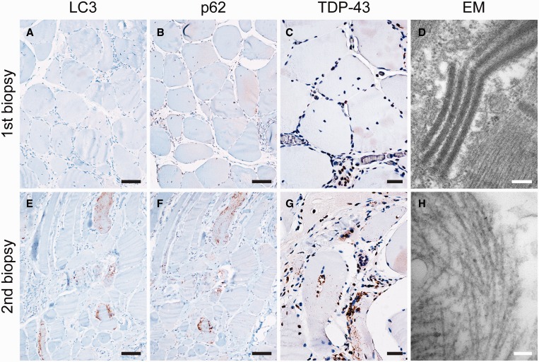
Immunohistochemical and ultrastructural analysis of repeat biopsies from patient #9. (A–D) The first biopsy at age 49 years demonstrated morphologic features of polymyositis (PM); (E–H) the second biopsy (7 years later) demonstrated morphologic features of inclusion body myositis (IBM). Immunohistochemical staining showed increased staining for LC3 ([E] vs [A]), p62 ([F] vs [B]), and TDP-43 ([G] vs [C]) in the second biopsy; the percentages of fibers positive for each marker are listed in Table 2. On ultrastructural analysis, the first biopsy showed mitochondrial crystalline arrays (D) but no 15- to 18-nm tubulofilamentous inclusions; these inclusions, diagnostic for IBM, were present in the second biopsy (H). Scale bars: A, B, E, F, 50 µm; C, G, 20 µm; D, 200 nm; H, 100 nm.

