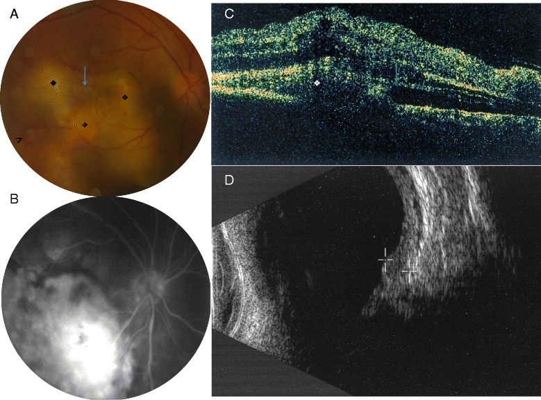
Fig. 1.

a Fundus examination of the right eye revealed a multifocal large yellowish choroidal infiltrate surrounding by hemorrhages (arrow head), associated with an overlying subretinal detachment, and subretinal fibrosis (arrow). b Fluorescein angiography showed late hyperfluorescence with focal vascular leakage. c OCT B scan showed choroidal infiltrates with serous retinal detachment and fibrosis (d) Ultrasound biomicroscopy confirmed the presence of a 3.8 mm parietal granuloma with few calcifications
