Figure 1.

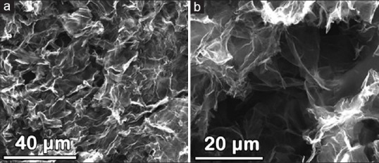
Scanning electron microscopic images of lyophilized graphene gels with (a) bar = 40 μm and (b) bar = 20 μm. Based on the images, the pore sizes range from hundreds of nanometers to several micrometers

Scanning electron microscopic images of lyophilized graphene gels with (a) bar = 40 μm and (b) bar = 20 μm. Based on the images, the pore sizes range from hundreds of nanometers to several micrometers