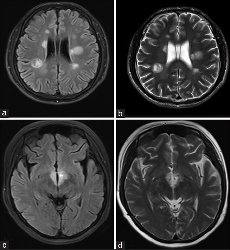
Figure 1.

Hallmark brain lesions for multiple sclerosis (a and b) and neuromyelitis optica spectrum disorder (c and d). (a) Axial fluid-attenuated inversion recovery sequence and (b) T2-weighted magnetic resonance imaging showed tumefactive multiple sclerosis lesions, presenting as a “fried egg” in periventricular areas. The yolk is the plaque itself and the white is the surrounding vasogenic edema. Typical neuromyelitis optica spectrum disorder lesions: (c) Fluid-attenuated inversion recovery sequence magnetic resonance imaging with (d) T2-weighted hyperintensities evident in periependymal areas.
