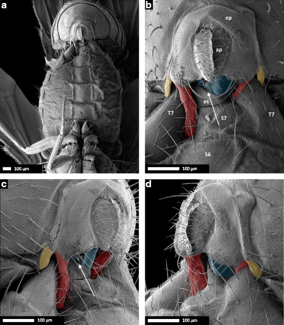
Fig. 5.

Scanning electron micrographs of coupled male and female D. pachea genitalia at 10 min after the beginning of copulation. a 37× and b 190× magnification of a ventral view, (c) left lateral view, (d) right lateral view. Male: ap, anal plates; ep, epandrium. Female: es, eversible sheath (intersegmental membrane between 7th sternite and oviscapt valves); S6, 6th sternite, S7, 7th sternite; T7, 7th tergite. The male’s epandrial lobes are artificially coloured in red, the male’s lateral epandrial spines in yellow and the female’s oviscapt valves in blue. The medial gap between the female’s oviscapt valves is indicated by an arrow and is visible from the left side, but not from the right side. Scale bar is 100 μm
