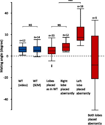
Fig. 6.

Comparison of the macroscale and microscale mating angles. Mating angles are shown for the wild-type (blue), with first the macroscale angle (based on video analysis) and second the microscale angle (based on SEM). Microscale angles are also presented for all treatments (left lobe shaved, right lobe shaved, left lobe cut, right lobe cut, symmetric mutant) combined together (red), but separated according to the positioning of the lobes. NS: not significant, ***: P < 0.001
