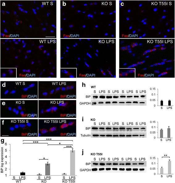
Fig. 6.

Systemic inflammation induces ER-stress most severely in the CNS of Cx32 KO T55I mutant mice. Images of brainstem sections focusing on white matter areas from saline (S) or LPS treated WT (a), Cx32 KO (b) and KO T55I (c) mice as indicated, immunostained with the ER-stress and pro-apoptotic marker Fas (red). Cell nuclei are stained with DAPI (blue). Higher magnification details of oligodendrocytes are shown in insets. Fas immunoreactivity is increased in LPS tissues, most prominently in KO T55I brainstem (c). Likewise, spinal cord longitudinal sections immunostained for the ER-stress marker BiP (red) (d–f) show increased immunoreactivity in LPS-treated mice compared to saline controls, especially in KO T55I mice. Scale bars in a-c: 30 μm; in d–f: 10 μm. g Real-time PCR analysis of BiP expression comparing LPS to saline groups from WT, KO and KO T55I animals, as indicated, shows that BiP mRNA is elevated in LPS tissues compared to saline controls in Cx32 KO and even more in KO T55I groups but not in WT mice. Direct comparison of LPS groups from all three genotypes confirms elevation of BiP expression in Cx32 KO and KO T55I mice compared to WT mice, and in KO T55I significantly more than in the simple KO group. h–j Immunoblot analysis of BiP levels (BiP specific band at 74 kDa) in brainstem tissue lysates (n = 4 mice per genotype and treatment group) from LPS compared to saline treated WT (h), KO (i) and KO T55 (j) mice as indicated, shows significant elevation of BiP levels only in the KO T55I LPS group. Blots were re-probed for tubulin or GAPDH for loading control and normalized BiP band intensity was quantified with Tinascan software (Student’s-test, *:p < 0.05, **:p < 0.01, ***:p < 0.001, Bonferroni corrected)
