Figure 9.
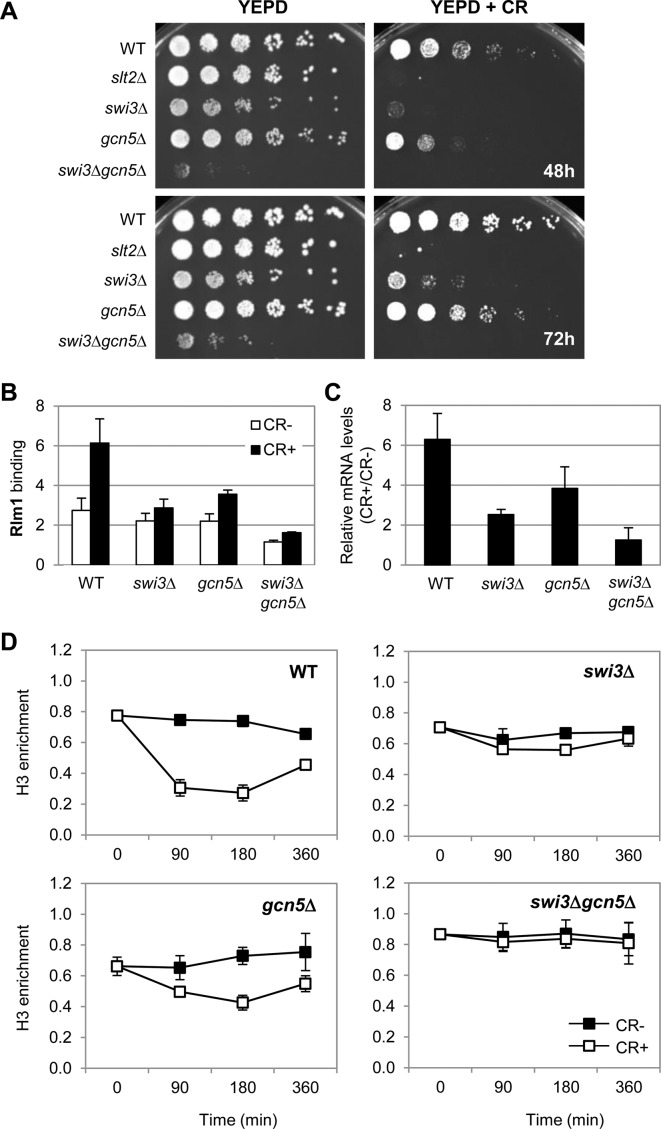
SWI/SNF and SAGA complexes cooperate to displace H3 histone at MLP1 and regulate gene transcription in response to cell wall stress. (A) The indicated strains were spotted on YEPD plates without or with 100 μg/ml of CR and plates were incubated for 48 and 72 h at 30°C. (B) Rlm1 enrichment at MLP1 promoter (MLP1-BOX1) was analyzed by ChIP in the indicated strains exposed to cell wall stress (CR 30 μg/ml, 3 h). (C) mRNA levels of MLP1 were analyzed by RT-qPCR in the same strains and growth conditions indicated in (A). Values represent the ratio between CR-treated and non-treated cells. Data represent the mean and standard deviation of at least three independent experiments. (D) The kinetics of histone H3 binding to MLP1 promoter (MLP1-BOX1) was determined by ChIP in WT, swi3Δ, gcn5Δ and swi3Δ gcn5Δ strains under CR treatment at the indicated times. ChIP assays were performed using an antibody against total H3. Data represent the mean and standard deviation of at least three independent experiments.

