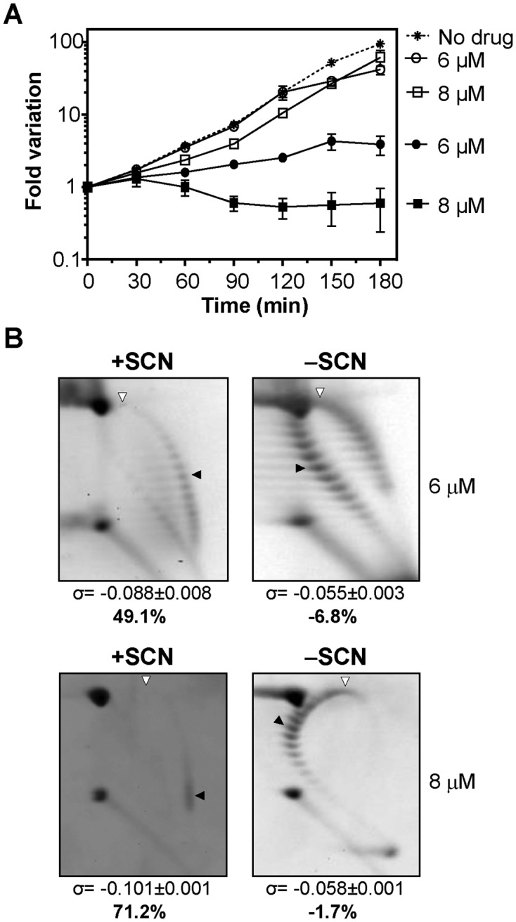
Figure 2.

The removal of SCN led to a recovery in the cell viability and in the supercoiling density. (A) Viability of R6 (pLS1) cultures. Cultures were grown at an OD620nm of 0.4 and diluted 100-fold in medium containing either 6 or 8 μM of SCN. After 120 min growth in the presence of the indicated amounts of SCN, cells were centrifuged, washed, and suspended in either drug-free medium (empty symbols) or in medium containing the same SCN concentrations (6 or 8 μM SCN, full symbols) than in the previous treatment. As control, a culture was grown at OD620nm of 0.4, diluted 100-fold in drug-free medium and grown during 180 min (no drug). Samples were taken at the indicated times and plated in drug-free agar medium. Results are the average of three independent replicates ± SEM. (B) Distribution of pLS1 topoisomers in 2D-agarose gel electrophoresis. Electrophoresis conditions and symbols are as indicated in the legend of Figure 1. Samples were taken after 120 min treatment with SCN (+SCN) and 120 min after removal of the drug (−SCN). Results are the average of at least three independent replicates ± SD.
