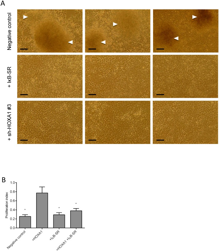
Figure 8.
Oncogenicity tests. (A) Foci formation assay was performed by transfecting MCF7 cells with empty vector (negative control), IκB-SR or sh-HOXA1 #3. After 2 weeks in culture, foci formation was observed for the negative control (white arrowheads) whereas cells transfected with IκB-SR or sh-HOXA1 did not form foci. Pictures presented were taken from three biologically independent experiments. Scale bar corresponds to 0.02 mm. (B) MTT assay was performed by transfection MCF10A cells with HOXA1 alone or in combination with IκB-SR. Global cell growth was determined by MTT assay. Results are presented as a proliferative index obtained by calculating the mean absorbance of each condition and corresponds to the mean of three biologically independent triplicates ± SEM. Statistical analysis was carried out using the ‘HOXA1’ condition as control level (*P < 0.0001).

