
Figure 1. Disruption of SPAK/OSR1 and cloning of a truncated isoform of OSR1 .
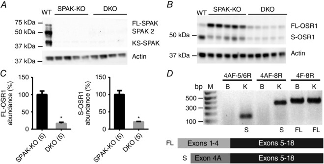
A, Western blot analysis of whole kidney lysates confirmed the absence of SPAK in SPAK knockout (SPAK‐KO) and SPAK/OSR1 double knockout (DKO) mice. B, Western blots showed a significant reduction in abundance of FL‐OSR1 and a short form of OSR1 (S‐OSR1) in SPAK/OSR1 DKO mice. C, quantification of FL‐OSR1 and S‐OSR1 abundance using blot in B. Densitometry values were normalized to actin and plotted as means ± SEM. FL‐OSR1: SPAK‐KO 100±10%, DKO 16±3%; * P = 6×10⁻⁵. S‐OSR1: SPAK‐KO: 100±12% and DKO: 21±1%; * P = 0.0001, unpaired t‐tests. D, 5′RACE PCR using a primer to exon 11 of OSR1 identified a transcript which lacks exons 1–4, and instead contains a novel first exon (4A), joined to exon 5 (see cartoon). This novel exon shares no homology with exon 5A of KS‐SPAK. RT‐PCR using total RNA from mouse brain and kidney revealed that this transcript may display tissue‐specific expression, as it is absent from brain. Numbers above bars indicate primer pairs used (see Methods). M, DNA ladder; bp, base pairs; B, brain; K, kidney; S, S‐OSR1 amplicon present; FL, FL‐OSR1 amplicon present.