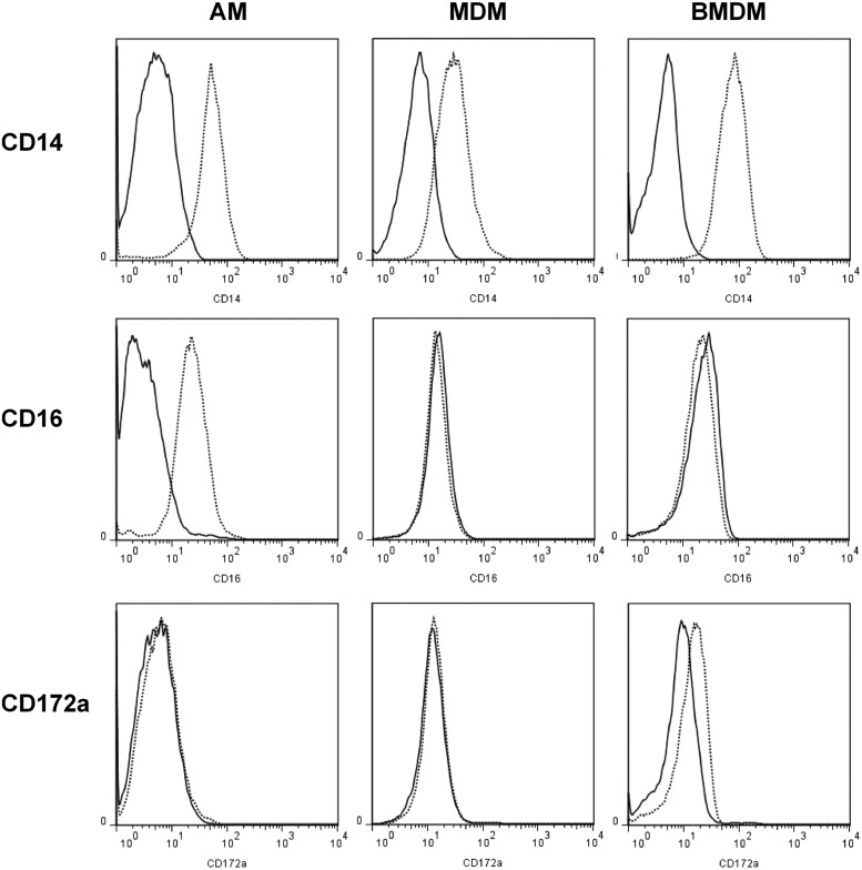
FIGURE 4.
Flow cytometry analysis of AMs, MDMs, and BMDMs from sheep. A lung lavage was performed to isolate AMs. Macrophages were differentiated from PBMCs and BM for 7 d with rhCSF1. Dead cells were excluded with propidium iodide. Solid lines represent isotype controls. Histograms are representative of three (AM and MDM) or five (BMDM) sheep.

