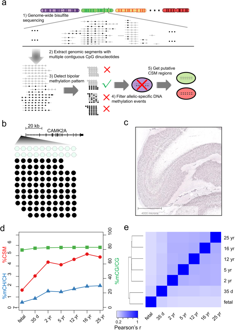
Figure 1. Identification of pCSM loci in human brain methylomes.
(a) Workflow for the computational inference of pCSM loci. (b) Methylation pattern of a predicted CSM loci (Chr5:149675541-149675604) at 6,138 bp upstream of CAMK2A gene in 25 yr brain methylome. (c) In situ hybridization for CAMK2A in human prefrontal cortex showing positive staining in the excitatory neurons in layers II-VI with absent staining in the glial cells (modified from Allen Brain Atlas; http://human.brain-map.org/ish/specimen/show/80936541?gene=811). (d) Changes of mC level in CG and CH context and the percentage of CSM segments after down-sampling normalization during human brain development. (e) Hierarchical clustering based on Pearson’s correlations of pCSM statuses predicted for 4-CpG segments in different human brain methylomes.

